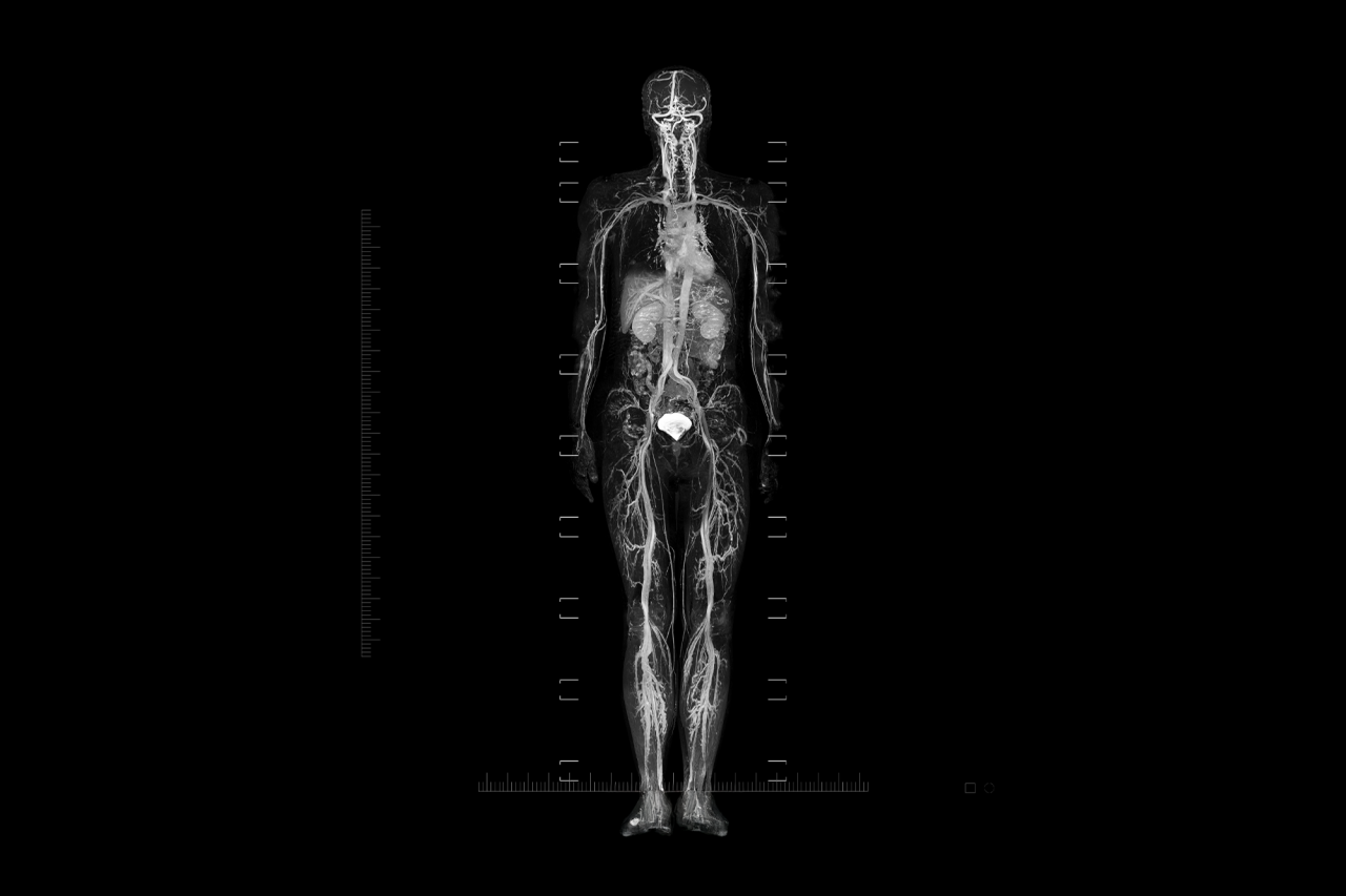
The pursuit of early cancer detection has driven remarkable innovation in medical imaging, and whole-body magnetic resonance imaging (WB-MRI) has emerged as one of the most discussed and debated tools in modern preventive medicine. Unlike traditional screening methods that target specific organs, WB-MRI offers a comprehensive evaluation of the entire body in a single session, promising to identify malignancies before symptoms manifest. This technology has captured public imagination, with direct-to-consumer imaging companies marketing full-body scans as a proactive approach to health management. However, the medical community remains divided on its appropriate role, particularly for asymptomatic individuals without elevated cancer risk.
This comprehensive analysis examines the scientific evidence, clinical applications, limitations, and future directions of whole-body MRI for cancer screening, providing healthcare professionals and informed patients with the nuanced understanding necessary to make evidence-based decisions.
Understanding Whole-Body MRI Technology
A. Fundamental Principles
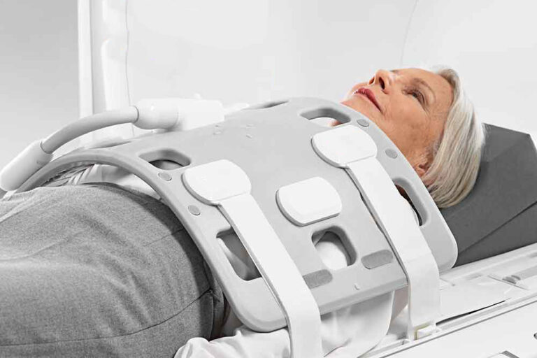
Whole-body MRI operates on the same foundational principles as conventional MRI, utilizing powerful magnetic fields and radio waves to generate detailed images of internal structures without exposing patients to ionizing radiation. Hydrogen atoms within body tissues align with the magnetic field, and radio frequency pulses temporarily disrupt this alignment. As the atoms return to their original state, they emit energy that sophisticated computer systems convert into high-resolution images of soft tissues, organs, and blood vessels.
The distinction between targeted MRI and whole-body MRI lies in scope and intent. A standard clinical MRI focuses on a specific anatomical region a knee, the brain, or the liver because clinical symptoms or history suggest pathology in that area. Whole-body MRI, conversely, stitches together imaging sequences from the top of the head to the mid-thigh (or occasionally to the toes) in a single session, typically lasting 40 to 60 minutes, usually without contrast dye.
B. Technical Protocols and Advancements
Contemporary WB-MRI protocols have evolved significantly, incorporating advanced sequences that enhance diagnostic capabilities. Diffusion-weighted imaging (DWI) has proven particularly valuable, providing functional insights that complement anatomical data and improve tumor detection sensitivity. This technique exploits differences in water molecule movement between normal and malignant tissues, where restricted diffusion in hypercellular tumor environments creates distinctive signal patterns.
The development of parallel imaging and accelerated acquisition protocols has reduced scan times while maintaining image quality, making the procedure more practical for clinical implementation. Large-field-of-view sequences enable comprehensive evaluation of the entire body while allowing concurrent detailed assessment of individual anatomical regions.
C. The ONCO-RADS Framework
A significant advancement in standardizing WB-MRI interpretation came with the development of the Oncologically Relevant Findings Reporting and Data System (ONCO-RADS). This standardized framework categorizes findings based on malignancy likelihood across five assessment categories, promoting consistency in acquisition, interpretation, and reporting. The system was developed by an international expert panel to enhance practice consistency and reduce variations that previously complicated multi-institutional collaboration.
Clinical Applications and Established Indications
A. Cancer Predisposition Syndromes
While WB-MRI’s role in general population screening remains controversial, its value in managing cancer predisposition syndromes is well-established. International guidelines now recommend whole-body MRI surveillance for individuals with conditions such as Li-Fraumeni syndrome, constitutional mismatch repair deficiency syndrome, and hereditary paraganglioma-pheochromocytoma.
Li-Fraumeni syndrome, caused by pathogenic germline variants in the TP53 tumor suppressor gene, confers extraordinarily high lifetime cancer risks approaching 100% by age 70. The SIGNIFIED study, a prospective observational investigation conducted in the United Kingdom, demonstrated that WB-MRI can detect early-stage cancers in this high-risk population, with 70% of detected cancers identified at stages amenable to curative treatment.
B. Multiple Myeloma and Metastatic Disease
WB-MRI has become an integral tool for staging and monitoring multiple myeloma, where the MY-RADS (Myeloma Response Assessment and Diagnosis System) framework provides standardized evaluation criteria. The technique’s superior soft tissue contrast enables detection of bone marrow infiltration and extraskeletal disease that may be missed by other imaging modalities.
Similarly, for prostate cancer staging, the MET-RADS-P (Metastasis Reporting and Data System for Prostate Cancer) has emerged as a reliable imaging biomarker for predicting metastatic disease progression and assessing treatment response.
C. Pediatric Applications
The use of WB-MRI in pediatric patients has expanded rapidly, particularly for screening children with cancer predisposition syndromes where the absence of ionizing radiation is advantageous given the anticipated requirement for numerous longitudinal examinations. Current indications include screening for presymptomatic lesions, tumor staging, investigating fevers of unknown origin, and monitoring rheumatologic and neuromuscular disorders.
Diagnostic Performance and Detection Rates
A. Sensitivity and Specificity Metrics
The diagnostic accuracy of WB-MRI varies considerably across studies and populations. A retrospective study examining WB-MRI surveillance in patients with cancer predisposition syndromes reported sensitivity of 64% and specificity of 92%, with a negative predictive value of 92%. These metrics suggest that a normal WB-MRI provides reasonable reassurance, though the moderate sensitivity indicates some cancers may be missed.
In pregnant patients, WB-MRI demonstrated 98% sensitivity and 88.5% specificity for cancer detection, leading researchers to describe it as “safe, efficient and the most effective method for detecting cancer” in this population.
B. Cancer Detection Yield
Meta-analyses of WB-MRI screening in asymptomatic individuals have reported pooled cancer detection rates ranging from 1.46% to 1.57%. While these percentages may appear modest, they translate to meaningful absolute numbers when applied to large screening populations. The Hercules study, a prospective real-world evaluation, aims to provide more definitive data on predictive accuracy and clinical utility using standardized ONCO-RADS and Clinical Significance Diagnosis frameworks.
C. Comparison with PET/CT
Whole-body MRI demonstrates comparable diagnostic accuracy to PET/CT for cancer detection while offering several advantages, including absence of ionizing radiation, no requirement for radiotracer injection, and lower costs. Studies comparing the two modalities for bone metastasis detection have shown strong concordance (κ = 0.87), with WB-MRI detecting 112 of 150 cases compared to PET/CT’s 115.
However, PET/CT retains advantages in certain applications, particularly for detecting small lung nodules and evaluating metabolic activity. For routine screening in asymptomatic individuals, WB-MRI’s radiation-free nature makes it preferable, as PET/CT’s high radiation dose renders it unsuitable for repeated use in healthy populations.
Critical Limitations and Risks
A. High Prevalence of Incidental Findings
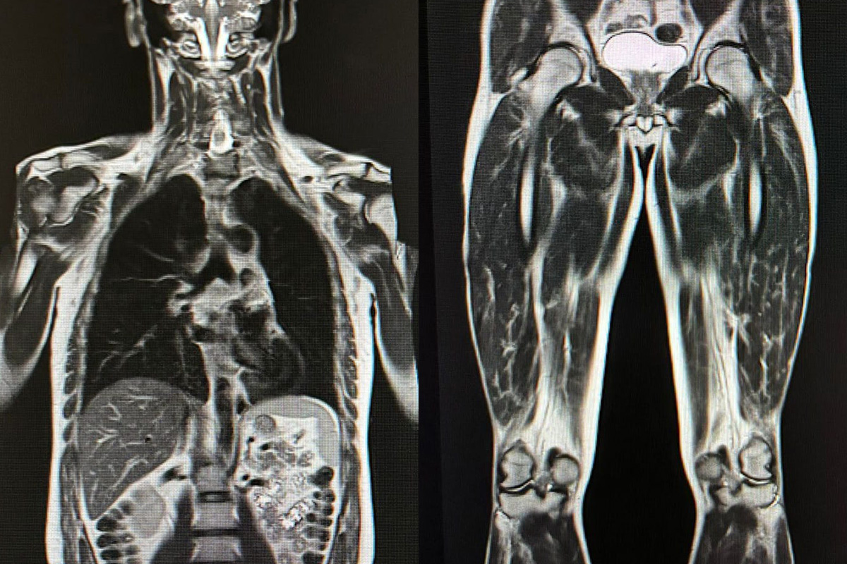
The most significant challenge facing WB-MRI screening is the extraordinarily high rate of incidental findings. Studies indicate that approximately 95% of screened patients have some abnormal finding, with roughly 30% having potentially relevant oncologic findings requiring further evaluation. While only about 1.8% of patients ultimately have confirmed malignancy, the vast majority of findings prove benign, creating substantial downstream consequences.
At the lesion level, meta-analyses have reported that 91% of abnormal findings are not relevant and considered benign, while 9% are potentially oncologically relevant and require further evaluation. This high prevalence of indeterminate findings generates considerable patient anxiety and drives unnecessary additional testing.
B. False Positives and Overdiagnosis
The low pretest probability problem fundamentally undermines screening effectiveness in average-risk populations. When the prevalence of serious disease is low, even highly specific tests generate substantial false-positive results. This statistical reality means that many healthy individuals undergo invasive procedures—biopsies, additional imaging, and occasionally surgery for findings that would never have caused harm.
The psychological burden of uncertain diagnoses should not be underestimated. Patients receiving indeterminate results may experience months of anxiety while awaiting follow-up examinations and definitive answers. The American Cancer Society notes that full-body scans can show “spots” or “shadows” that could be serious but are often normal variations, creating stress without proportional benefit.
C. Limited Evidence for Mortality Benefit
Perhaps most critically, no randomized controlled trials have demonstrated that WB-MRI screening extends lives or improves quality of life in average-risk populations. The available evidence primarily addresses exam performance metrics sensitivity, specificity, and cancer yield rather than long-term patient outcomes.
The Fred Hutchinson Cancer Center emphasizes that an effective screening test must detect cancer at a preclinical stage and confer a survival benefit compared to diagnosis based on clinical symptoms. While WB-MRI is minimally invasive, it is neither low-cost nor proven to provide survival benefits.
D. Blind Spots and Limitations
WB-MRI has notable limitations in certain anatomical regions. Small lung nodules may be missed, as the technique is less sensitive than low-dose CT for pulmonary pathology. Colon polyps and breast microcalcifications targets of established screening programs may also escape detection. These blind spots overlap with areas where proven screening methods excel, reinforcing the importance of continuing guideline-recommended screenings regardless of WB-MRI results.
Economic Considerations and Access
A. Cost and Insurance Coverage
Consumer full-body MRI scans typically cost between $1,000 and $3,000 out of pocket, with commercial providers such as Prenuvo charging approximately $2,499 for comprehensive scans. Insurance companies rarely cover WB-MRI for general screening or wellness purposes, as most commercial and government plans do not recognize it as medically necessary for asymptomatic individuals.
This cost barrier raises significant equity concerns. The Hercules study has incorporated a health equity arm, offering subsidized access to reduce financial barriers for underserved populations with sliding-scale subsidies based on socioeconomic factors.
B. Cost-Effectiveness Questions
Formal cost-effectiveness analyses of WB-MRI screening remain lacking. The downstream costs of investigating incidental findings—additional imaging, biopsies, specialist consultations, and in some cases unnecessary treatments—add substantially to the direct screening costs. Without evidence of mortality benefit, these expenditures cannot be justified on a population health basis.
Current Guidelines and Recommendations
A. Professional Society Positions
Major medical organizations have adopted cautious positions regarding WB-MRI screening in average-risk populations. The Royal Australian and New Zealand College of Radiologists (RANZCR) explicitly does not recommend whole-body MRI screening in asymptomatic patients who lack a previously diagnosed malignancy or cancer predisposition syndrome.
Similarly, the American College of Radiology has stated that elective MRI screenings are not recommended and may cause more harm than good, emphasizing the absence of evidence that such screening extends or improves quality of life.
B. Evidence-Based Indications
Current guidelines support WB-MRI for specific high-risk populations:
A. Individuals with Li-Fraumeni syndrome and other TP53-related cancer predisposition syndromes
B. Patients with constitutional mismatch repair deficiency syndrome
C. Those with hereditary paraganglioma-pheochromocytoma syndromes
D. Patients undergoing surveillance for multiple myeloma
E. Men with advanced prostate cancer requiring staging and treatment response assessment
For these populations, the balance of benefits and risks favors regular WB-MRI surveillance, with evidence demonstrating detection of early-stage, potentially curable cancers.
Future Directions and Emerging Technologies
A. Artificial Intelligence Integration
The application of deep learning methods to WB-MRI holds promise for improving diagnostic accuracy and reducing radiologist workload. Artificial intelligence algorithms may enhance lesion detection, characterize indeterminate findings more precisely, and reduce false-positive rates that currently drive unnecessary downstream testing.
B. Liquid Biopsy Integration
Combining WB-MRI with multi-cancer early detection (MCED) blood tests represents another promising avenue. Preliminary research suggests that integrating imaging findings with circulating tumor DNA analysis may improve diagnostic specificity and reduce the psychological burden of indeterminate results.
C. Protocol Standardization
Ongoing efforts to standardize WB-MRI protocols across institutions aim to reduce variability that currently complicates multi-institutional collaboration and care standardization. The ONCO-RADS framework represents a significant step toward consistent acquisition, interpretation, and reporting practices.
Practical Guidance for Patients and Clinicians
A. When to Consider WB-MRI
Whole-body MRI may be appropriate for:
A. Individuals with confirmed cancer predisposition syndromes requiring surveillance
B. Patients with multiple myeloma or certain metastatic cancers requiring staging
C. Cases where conventional imaging has been inconclusive despite clinical suspicion
D. Research participants enrolled in IRB-approved studies evaluating screening efficacy
B. When to Exercise Caution
WB-MRI is generally not recommended for:
A. Average-risk asymptomatic adults without specific indications
B. Patients seeking general health screening without medical justification
C. Individuals who have not completed evidence-based organ-specific screenings
D. Those unable to tolerate the procedure or with MRI contraindications
C. Shared Decision-Making
Patients considering WB-MRI should engage in thorough discussions with their healthcare providers, addressing:
A. The limitations of available evidence regarding mortality benefit
B. The high likelihood of incidental findings requiring follow-up
C. The out-of-pocket costs and lack of insurance coverage
D. The importance of continuing established screening programs
Conclusion
Whole-body MRI represents a powerful imaging technology with established value in specific clinical contexts, particularly for cancer predisposition syndromes and oncologic staging. However, its role as a general screening tool for asymptomatic populations remains unsupported by current evidence. The high prevalence of incidental findings, substantial false-positive rates, uncertain mortality benefits, and significant costs create an unfavorable balance for average-risk individuals.
The technology continues to evolve, with advances in diffusion-weighted imaging, artificial intelligence, and integration with liquid biopsies potentially addressing current limitations. Until randomized controlled trials demonstrate meaningful improvements in patient outcomes, WB-MRI should be reserved for evidence-based indications under appropriate clinical supervision. Patients deserve honest, nuanced information about what this technology can and cannot deliver not marketing promises that outpace scientific reality.
For those with elevated cancer risk due to genetic predisposition or other factors, WB-MRI offers genuine value within comprehensive surveillance programs. For the broader population, the path to reducing cancer mortality lies not in indiscriminate whole-body scanning but in adherence to proven screening methods, attention to warning symptoms, and continued investment in research that will ultimately determine whether this promising technology can fulfill its theoretical potential.